
L-spine surgery
Percutaneous Laser Disk Decompression (Sclerotherapy)
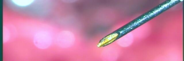
With the help of an X-ray the surgeon inserts a thin needle into the intervertebral disk. Due to the administration of analgetics the entire intervention is nearly pain free. By the help of the needle the surgeon can then insert a glass fiber which carries a laser. The slipped disk can then be vaporized by a laser.
Microsurgery
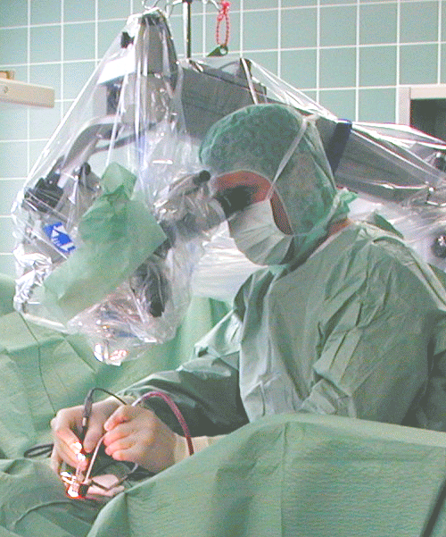
Similar to an operation of the cervical spine it is possible that the spinal nerves get comprimised by either a slipped disk or a narrowing of the spinal canal caused by an osseous deformity. Depending on the severity of the discomfort it is sometimes not possible to treat the patient without an operation. It is always important to distinguish between a nerval irritation or a nerval damage which can lead to paralysis, loss of sensation or bladder- colon disturbances. All microsurgical operations get performed with a general anesthesia.
A couple of days before the microsurgical intervention the patient needs to introduce himself at the hospital. This procedure makes sense because you get to know the anesthetist and the nursing service. For the operation the patient comes to the hospital at the same morning as the operation is scheduled.
Procedure:
During the operation the surgeon will make a 3-4 cm long cut according to the operation segment. Afterwards the muscular structures get moved aside and ligaments as well as bony structures of the vertebral bodies get removed to open the vertebral canal. The dissection of the ligamental and bony structures is necessary because this also releases a lot of stress from the nerval roots. Following this the surgeon prepares the chamber of the intervertebral disk. In the end the cut gets sutured level by level.
If it comes to a pronounced slipped disk the surgeon will dissect free disk tissue. This is only the case if the available disk tissue is not usable anymore. In most cases the surgeon tries to obtain most of the original structures.
After the operation
At the day of the operation it is already possible to stand up if you are in company. It is advisable to avoid sitting and walking for a longer period during the following 8 days but it is definitely more helpful to stand up for a short period a couple of times per day in place of standing for longer periods. In most of the cases it is possible to release the patient from the hospital after 1- 4 days after the operation. Before this the patient gets advice from remedial gymnastics which helps to learn special movements which prevents the vertebral column from further damage. The stiches must not be taken out and the patient maximal capacity can be restored after 6-8 weeks under ideal circumstances.
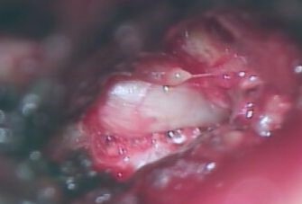
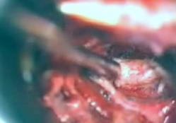
durch einen Bandscheibenvorfall verdrängte Nervenwurzel, Blick durch ein OP Mikroskop
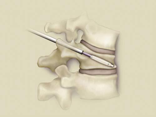
Kyphoplasty
The Kyphoplasty can be seen as a minimally invasive procedure to treat a vertebral fracture. During the operation the surgeon will introduce bone cement into the fractured vertebra via a thin canula. The patient can stand up directly after the procedure and experiences a direct alleviation of pain in most of the cases.
The indication for a kyphoplasty is given when the patient suffers from a stable fracture which he can experience during an accident or due to osteoporosis.
Advantages:
Minimally invasive – low operational stress – save – easy and fast mobilization of the patient after the operation – very effective, especially for pain treatment
|
|
|
|
|
Insertion of the baloon |
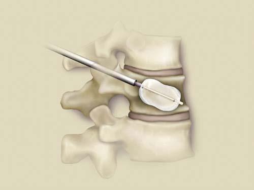
Inflation |
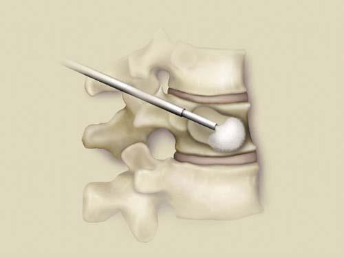
Filling with bone cement |
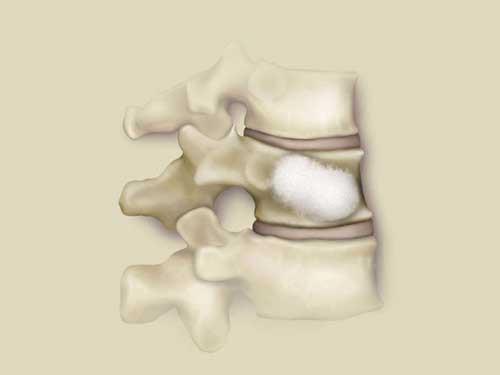
Filled and stabalised vertebra |